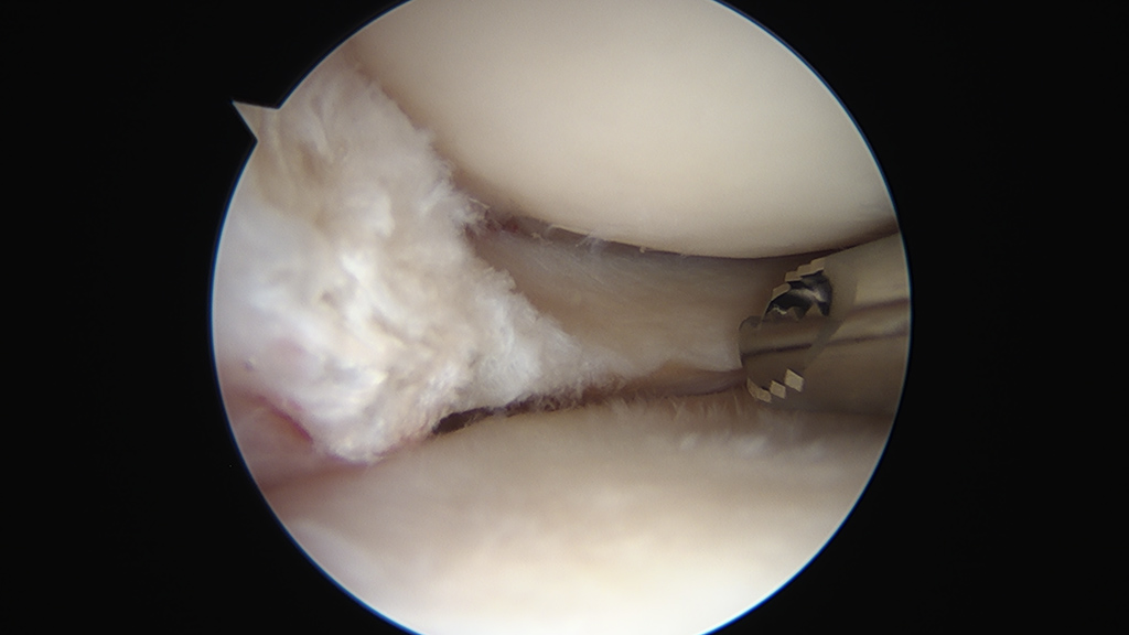
The knee has two c-shaped cartilages called ‘menisci.’ These function as ‘shock-absorbers’ within the knee, distributing load within the joint. The menisci can be torn as the result of pivoting injuries or as part of long-term wear. Sometimes these tears may cause ongoing pain or persisting mechanical catching within the knee. In such cases the tear may be surgically managed with knee arthroscopy. This is a ‘key-hole’ procedure in which small incisions are used to introduce a camera and a working instrument into the knee in order to address the tear. The use of key-hole surgery means that patients generally have a rapid recovery and can usually return home the same day as their surgery.